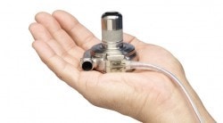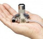
This surgeon’s team has previously done work using mechanical engineering technology to help them make better decisions, you may have read about their use of 3D Printing to evaluate different treatment options. They often work with computer models of patients and devices n collaboration with spinal surgeon Dr. Sandro LaRocca in New Jersey, so they had almost all the tools they needed to help this patient.
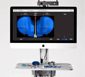
For this case, they had a computer model of the smaller assist device, and a computer model of the patient’s heart area that they extracted from a CAT scan. Using those two models and visualization software they were able to insert the device model into the body model to verify that the smaller device would fit.
The issue they faced was that they had no computer model for the larger device. Creating a model the traditional way would take to long. So they called PADT and asked if we could scan the actual object and give them a computer model that they could use.
Just in Time Scanning
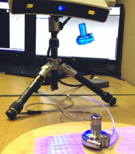
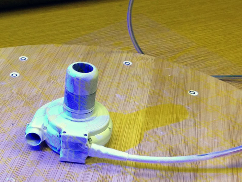
One of PADT’s engineer, Johnathon Wright, took the device to our Geomagic Capture blue light scanner to extract a surface model from the real part. In this image you can see the device being scanned:
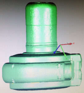
In this image you can see what the part looks like to the scanner: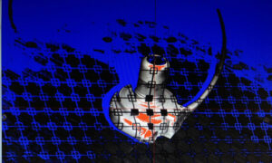
Here is what the point cloud looks like when the scan is completed:
In about an hour, Johnathon was able to go from “can you do this” to a water-tight solid that the Doctor could use with his computer model of the patient to see if this larger, better part fit in the patient’s chest.
Here is what the whole setup looks like:
Johnathon used Geomagic’s scanning tools running on a PADT CUBE computer that is specifically optimized for scanning to make the process faster and more accurate. In the past, a task like this would have required an expensive and temperamental laser scanner, a dedicated lab, and probably four to eight hours of engineering time to clean up the resulting scan data. As you can see, the device sits on a desktop and requires very little infrastructure or special equipment.
Disruptive Technology
Any day we can help a physician strive for a better surgical outcome is a good day. Beyond that this is also a great example of how three important aspects of the technology enabled us to deliver useful information quickly, making desktop scanning a disruptive technology.
The first key technology is the blue-light scanning itself. A form of structure-light 3D scanning, this approach uses a blue light because it contrasts the object better. The breakthrough with this technology is that it does not require expensive lasers or complex optics. Faster computing allows for the complex algorithms used to be quickly and accurately applied. The approach does not require any special equipment beyond the scanner itself. This results in an affordable device that is easily deployed and operated. How easy, the 3D motion capture device on the Microsoft Xbox Kinect is a structure-light 3D scanner – using infrared light instead of blue.
Modern software used to convert the scan data into useful information is the second technology deployed for this solution. In the past the process of calculating the points on a scanned surface, cleaning up spurious data, and converting it to a form that could be easily used was tedious and difficult. The Geomagic software suite has a modern, intuitive user interface that sits on top of very sophisticated tools that automate many of the steps that used to take us hours to carry out.
The final key technology that makes desktop scanning so disruptive is one that we take for grated today: standards. We were able to produce an STL file from the scan data and the Doctor’s team was able to read that directly in to their visualization software. It is a simple thing, but without standard file formats, transferring so much data would also involve translators which introduce errors and time.
Engineering Better Outcomes
Here at PADT we truly enjoy applying technology developed in the Aerospace or electronics space to other industries, especially medical applications. This is another great example of how useful engineering tools can be, improving someones life directly.

